BNA Annual General Meeting 2025
1st April 2025
Below you can find the abstracts for the talks during our BNA Members Meeting. Click here for more information about the programme, registration, and other details about the Members Meeting.
Hunting(tin) for Genetic interactions in Huntington's Disease
 Rachel Sellick, PhD researcher, Cardiff University
Rachel Sellick is a final Year Phd student at Cardiff University. After studying her BSc(Hons) in Anatomy and an MRes in Translational Medicine, Rachel became increasingly interested in the complex nature of the brain, and how we can slow progression in sufferers of neurological and neurodegenerative disease. After an 18 month stint as a Research Assistant at Cardiff University, Rachel was accepted onto the 4 year SWBio Doctoral Training Partnership and under the supervision of Dr Michael Taylor, Professor Anne Rosser and Dr Mariah Lelos, is investigating protein and genetic interactions associated with Huntingtin. Subsequently, Rachel is interested in how manipulation of various genes and proteins can slow symptom onset in Huntington's Disease. When Rachel isn't studying, she is passionate about sharing her research through communication and marketing, runs her own music business (Scarlet River PR) and hits the hockey field as an Umpire.
Abstract
Medium Spiny Neuron (MSN) degeneration is a major pathological hallmark in the genetically inherited, Huntington’s Disease (HD) for which there is no cure or treatment. Mechanisms associated with neurodegeneration in HD is thought to be associated with protein-protein and genetic interactions that vastly interfere with cellular functions and networks via its interaction with mutant Huntingtin (mHTT). Understanding these mechanisms is fundamental in providing insight into causes of neuronal loss and degeneration and, may aid in the development of new therapeutics to slow progression and enhance quality of life. FoxP1 and Mef2C are transcription factors known to play a role in tissue- and cell-specific regulation, and striatal development. Previous data has shown FoxP1 and Mef2C to interact with HTT, and potential manipulation of the gene is able to rescue a neurodegenerative phenotype. This PhD project has used Drosophila melanogaster and mouse models of HD to investigate the effects of polyglutamine repeat expansion and the effect of overexpression and downregulation of genes on neurodevelopment and degeneration in models of HD.
Investigation of COMP360 effects on neuronal ensembles using FosTRAP system in mice.
 Nutthaya Bunmak, Postgraduate, University of Bristol
2023 – Present: PhD student in Neuroscience at the University of Bristol, Dissertation: Mapping the effects of COMP360 psilocybin on neuronal ensembles using FosTRAP mice
2019 – 2021: Mahidol University, Thailand - M.Sc. Physiology, Faculty of Science [First class honour]; Dissertation: "The Correlation of Growth Factors and Cytokines with Cognitive Function as Biomarkers for Cognitive Impairment in Chronic Medicated Schizophrenia"
2015 – 2018: Mahidol University, Thailand - B.Sc. Biotechnology, Faculty of Science [First class honour]; Senior Project: "Co-culture of immortalized-Hepatocyte Cells with Fibroblasts to Improve Proliferation and Hepatic Functions As a Drug Screening Model"
Publications:
Neuropsychopharmacology Reports (2022) - "Correlation of BDNF, VEGF, TNF-α, and S100B with cognitive impairments in chronic,medicated schizophrenia patients", DOI: 10.1002/npr2.12261
The 25th Thai Neuroscience Society International Conference 2022 - "Correlation of BDNF, VEGF, TNF-α, and S100B with cognitive impairments in chronic, medicated schizophrenia patients"
Conference The 24th Thai Neuroscience Society International Conference 2021 - "Glial derived neurotrophic factor (GDNF): a potential biomarker for cognitive deficit in chronic schizophrenia"
The 20th Science Project Exhibition 2018 - "Co-culture of immortalized-Hepatocyte Cells with Fibroblast to Improve Proliferation and Hepatic Function"
Abstract
Psilocybin has been widely tested for multipurposes in treating neuropsychiatric disorders. Several studies have demonstrated the single-dose psilocybin potential to reorganize neural networks, improve neuroplasticity, induce positive affective states and alleviate depressive and anxiety symptoms. However, psilocybin has not yet been applied in clinics because of the inconclusive mechanism behind its therapeutic properties and long-lasting effect, the requirement of psychotherapeutic support during treatment sessions, and the scarcity of results from repeated treatments.
In this study, we aim to reveal the target brain regions activated by repeated doses of the COMP360 psilocybin, under neutral and manipulated affective states mice. Using the genetically altered mice, Fos2ACreERxtdTomato, we can permanently label the neuronal cells across multiple regions activated by psilocybin. The neurons in the target regions will be further investigated on their activities and connections to explain how repeated doses of COMP360 psilocybin act on the brain for its therapeutic functions. This study could fulfil the understanding of the COMP360 psilocybin mechanism and provide benefits towards the development of potential therapeutic interventions for neuropsychiatric treatments.
Underlying Neural Mechanisms of Working Memory in Older Adults
 Esteban León Correa, PhD student, Edge Hill University
Hello! My name is Esteban Leon Correa, originally from Ecuador, and I am currently in the final year of my PhD journey at Edge Hill University. My research project has centred on understanding neural functioning in older adults with a focus on topics such as brain modulation, cognitive training, and cognitive load. I employ a wide variety of methodologies including EEG, tDCS, TMS, statistical modelling and machine learning. I firmly believe in the transformative potential of science to positively impact society. Thus, I am particularly drawn to projects that tackle tangible real-world challenges.
Abstract
According to the World Population Ageing report of the United Nations (UN) the number of older adults (aged 65 or over) will be more than doubled within the next 30 years. Ageing is characterised by a progressive neurodegeneration that affects cognitive skills like working memory (WM) that are crucial for daily living. While structural changes in the old brain associated with impairments in WM are widely known (e.g., loss of grey in hippocampus), functional changes and compensatory mechanisms in this population remain unclear. In this talk I will present a group of studies that we conducted where we tried to address the aforementioned topics. In the first study, we explored the role of the prefrontal cortex in WM in older adults using a combination of transcranial magnetic stimulation (TMS) and electroencephalography (EEG). In the second study, we compared the brain functioning of younger and older populations using an EEG while they were completing challenging WM tasks. And in the last study we tested the potential benefits of cognitive training paired with transcranial direct current stimulation (tDCS) in older adults in enhancing WM and brain functioning. We will discuss some of the preliminary findings and potential future directions.
Study of oligodendrocytes and myelination during development in a rat model of fragile X syndrome
 Eleni Tsoukala, PhD student, University of Edinburgh
My name is Eleni Tsoukala and I am a second year PhD student at the Zoupi lab at the University of Edinburgh. I obtained my undergraduate degree in Biology from the National and Kapodistrian University of Athens. For my undergraduate thesis, I worked with mouse models of multiple sclerosis, and this is when I became fascinated with oligodendrocytes and myelin. Then I moved to Edinburgh, where I completed my master’s degree in Integrative neuroscience, and became interested in neurodevelopmental disorders. So, I joined the SIDB PhD program and the Zoupi lab to investigate how oligodendrocytes contribute to the physiology of fragile X syndrome, a leading syndromic neurodevelopmental disorder.
Abstract
Fragile X syndrome (FXS) is one of the leading forms of syndromic autism spectrum disorders (ASDs), caused by loss of Fragile X messenger ribonucleoprotein (FMRP). In the central nervous system (CNS), FMRP is expressed by neurons and all types of glial cells. Oligodendrocytes are the glial cells that produce myelin, a membrane that wraps the axon and speeds up signal transmission. The process of myelin production is called myelination, and it occurs primarily after birth in the CNS. Oligodendrocyte and/or myelin alterations have been reported in human and animal studies of FXS but their role in FXS pathophysiology remains unknown. We hypothesize that myelination is necessary for neuronal circuit function. To test our hypothesis, we use the rat model of FXS [Fmr1 knockout (KO) rats and their wild type (WT) littermates]. In vitro data from our lab show decreased oligodendrocyte maturation in Fmr1 KO rats, compared to their WT littermates. This is corroborated by preliminary in vivo data, which suggests that myelin sheath production is delayed in Fmr1 KO rats compared to their WT littermates. Future experiments will assess how these changes affect the speed of signal transmission and if manipulations of myelination are beneficial for cellular and functional recovery.
 Fengxi Jin, Postgraduate, The University of Manchester
Fengxi Jin, a PhD student at the University of Manchester and a member of the British Neuroscience Association (BNA), has embarked on a unique journey in neuroscience that skillfully blends his background in mathematics with a focus on the visual-guided behavior of freely moving mice. His aim is to elucidate the complex interplay between arousal and movements on neural activity within the visual pathway, addressing a long-standing scientific challenge. His research is organized into two main projects.
In the first project, Fengxi leverages 3D software to simulate mouse vision, investigating how visual stimuli affect their behavior. This technology enables highly accurate simulations of mouse vision.
The second project involves the use of a miniaturized endoscopic camera, along with head/body tracking, eye tracking, and a custom-designed vestibular platform. This setup allows for the meticulous manipulation and observation of arousal and movement in real-time.
Abstract
Arousal and movements affect neural activity along the visual pathway. These two variables are correlated during behaviour, and we don’t have a systematic way to determine their relative contributions to sensory processing.
To overcome this problem, I developed a system to measure arousal as pupil constriction in freely moving animals. The system is based on a miniaturised endoscopic camera that can be stably attached to the animal head during experiments. I then developed an experimental protocol to separate the effects of arousal, passive and active body movements by combining head/body tracking, eye tracking and a custom-designed vestibular platform.
Results: I will present behavioural results in which we can separate arousal and movement effects by changing the frequency of vestibular stimulation obtained by tilting the platform. I will also show some preliminary results on the impact of movements on visual evoked sensations in the primary visual thalamus.
The system I developed enables the measurement of arousal in freely moving animals and can be used to quantify the effects of arousal on neural activity.
Role of Primary cilia and the DNA damage response in the aetiology of Motor neuron disease
 Vasanta Subramanian, Senior researcher/Clinician, University of Bath
Vasanta Subramanian did her undergraduate and PhD studies from the University of Delhi and the Indian Institute of Science , both of which are India’s premier research Institutions. After Post-doctoral stints at the ICRF-DBU in Oxford and the Max Planck Institute for Biophysical Chemistry in Gottingen, Germany, she joined the University of Bath where she currently a Reader in Vertebrate Developmental Genetics.
Vasanta’s research focuses on neurodevelopmental and neurodegenerative disorders. She uses a broad range of cell and molecular biology techniques, 3D organoid culture, CRISPR/Cas9 technology and developmental genetics involving transgenic and knockout mouse models as well as the rapidly aging killifish model to understand disease mechanisms.
She is deeply committed to EDI issues and is currently the BNA’s EDI representative. In addition to training and mentoring her own research group, over the years she has hosted a large number of school and undergraduate students on externally funded summer bursaries to encourage them to consider a career in science.
Besides her research, Vasanta enjoys travelling, photography and Indian Classical music.
Abstract
Mutations in CFAP410, a basal body protein known to be required for the formation of primary cilia, have been recently identified in amyotrophic lateral sclerosis (ALS), a devastating late onset neurodegenerative disorder with poor prognosis. CFAP410 is also implicated in the DNA damage response and interacts with Nek1 also shown to be mutated in ALS. In this study we have investigated the effect of knocking in a HA epitope tag and two ALS associated mutations into the endogenous Cfap410 gene by gene editing using CRISPR/Cas9 in mouse embryonic stem cells (mESCs). We show that primary cilia in these mESCs, the neural progenitors and neurons differentiated from these mESCs do not exhibit any gross abnormalities. However, ESCs, neural progenitors and neurons with knockin Cfap410 ALS variants are more susceptible to DNA damage and exhibit impaired interaction with Nek1. Our findings point to DNA damage as a convergent pathway leading to ALS.
The ventral hippocampus and reversal learning
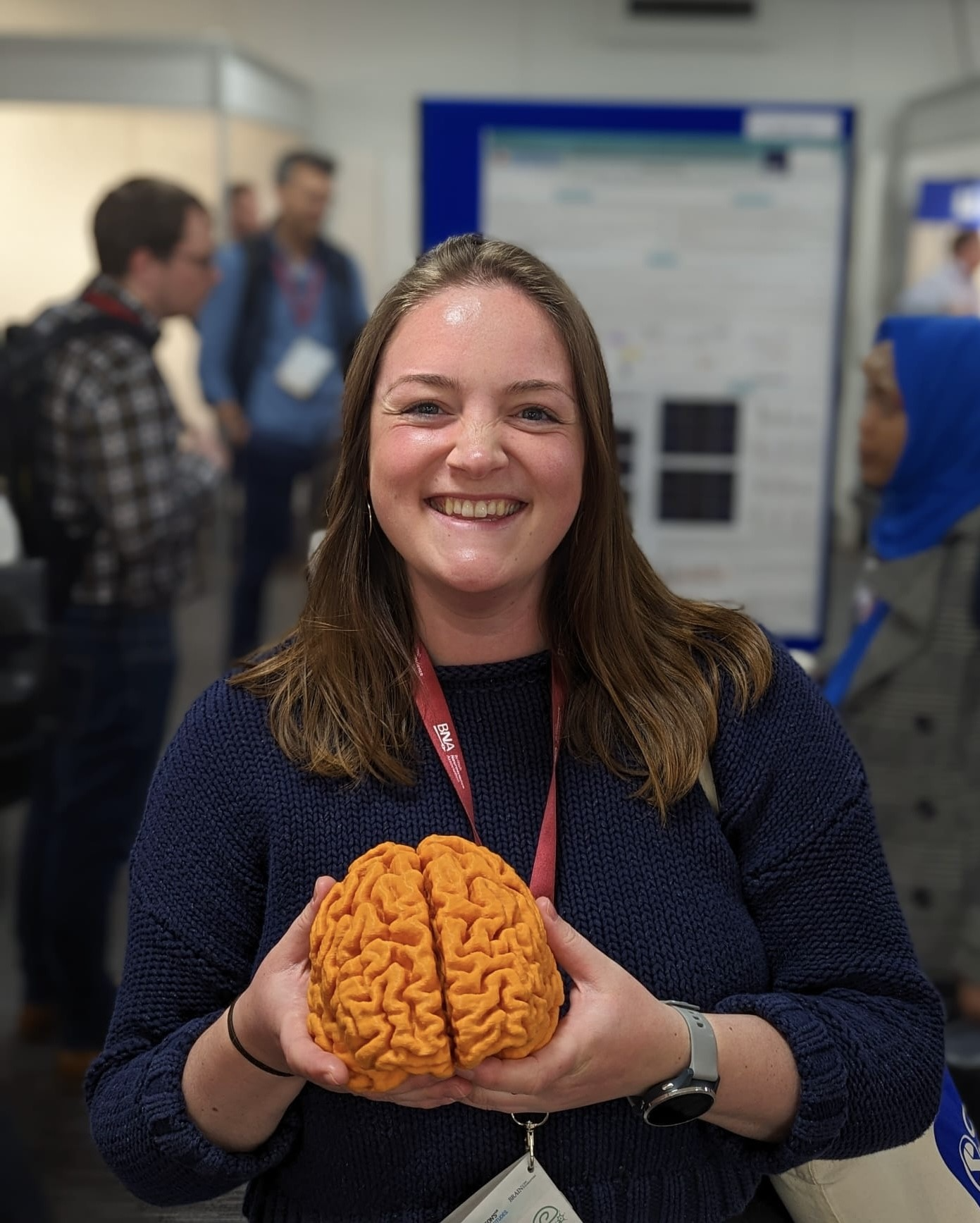 Rachel Grasmeder Allen, PhD Student, University of Nottingham
Rachel is a third year BBSRC funded PhD student in the School of Psychology at the University of Nottingham. Her research uses in vivo models to investigate the role of the hippocampus in cognitive and behavioural functions. She is specifically interested in GABAergic inhibition and disinhibition in the ventral hippocampus and how altering activity levels in the ventral hippocampus can affect distinct stages of reversal learning.
Abstract
Reversal learning, a form of cognitive flexibility disrupted in many brain disorders, involves switching from one response to another when the reward contingencies of the responses are reversed. The hippocampus, especially the ventral hippocampus (VH), connects to fronto-striatal circuits regulating reversal learning. Previous studies reported hippocampal lesions impaired, whereas chemogenetic activation of VH facilitated, the acquisition of reversal learning in animal models. However, we still have limited evidence on how VH activity affects distinct stages of reversal learning.
Therefore, we examined the impact of VH functional inhibition or disinhibition, by infusion of a GABAA receptor agonist (muscimol) and antagonist (picrotoxin), on repeated reversal learning and acquisition of reversal learning in male Lister hooded rats performing a two-lever discrimination task.
VH functional inhibition, but not disinhibition, impaired repeated reversal learning, indicating that repeated reversal learning requires VH activity, but not balanced levels of VH activity. In contrast, VH disinhibition, but not inhibition, impaired expression of the previous response during reminder trials preceding reversal trials, suggesting such expression does not require VH activity but may be disrupted by aberrant activation of projection sites. Our ongoing research is testing whether VH has a role during the acquisition of reversal learning.
Inflammation as a potential contributor to depression in people living with HIV
 Arish Mudra Rakshasa-Loots, PhD Researcher, University of Edinburgh
Arish Mudra Rakshasa-Loots (he/him) is a neuroscientist interested in improving mental health outcomes amongst people living with HIV. He is currently completing a PhD in Translational Neuroscience at the University of Edinburgh.
Abstract
People living with HIV are three times more likely to experience depressive symptoms compared to people without HIV. HIV elicits inflammation in the central nervous system and periphery, which persists despite antiretroviral therapy.
We explored the possible role of inflammation in driving the risk for depression in this community. First, we tested whether key inflammatory biomarkers mediated the risk for depressive symptoms in people with HIV. Amongst older adults (n = 204), controlling for the effects of MIG and TNF-α in plasma and MIP1-α and IL-6 in CSF resulted in >10% attenuation of the odds ratio for depressive symptoms in people with HIV, suggesting that these biomarkers may be potential mediators of these odds. Next, we used diffusion-weighted magnetic resonance spectroscopy to show that the severity of depressive symptoms in people with HIV (n = 16) was strongly correlated with intracellular diffusion of the neuroinflammatory biomarkers creatine (rho = 0.72) and choline (rho = 0.58) in the anterior cingulate cortex.
Together, these findings suggest that inflammation in the brain and the body may contribute to depressive symptom severity in people with HIV. With further validation, this work may inform early detection and individualised intervention for depression in people with HIV.
 Matthew Rowan, Assistant Professor, Emory University
Dr. Matthew Rowan is an Assistant Professor in the Cell Biology department at Emory University. The Rowan lab is interested in cellular and molecular mechanisms of different neuron types which allow these incredibly diverse cells to perform the wide variety of signaling functions necessary for learning and cognition in the mature mammalian forebrain. We recently have focused on how physiological and synaptic vulnerabilities arise depending on cortical region or in particular neuronal subtypes. We employ a multifaceted, intersectional approach to perform these studies in intact circuits from mature brains, using 2P optical imaging, AAVs, patch clamp electrophysiology, chemogenetics, CRISPR, and modeling techniques, in in vivo and ex vivo experiments. Using these approaches, we aim to uncover changes in different cortical regions and neuronal cell classes. We strive to better understand molecular mechanisms of excitability and plasticity of neurons, and how these properties contribute to the functional operation of local circuits as a whole, in both healthy and disease contexts.
Abstract
Although the dendrites of L2/3 PCs are impressive, they lack the obvious anatomical compartments of their layer 5 PC counterparts. For example, many L2 PCs do not display a clear distal ‘tuft’ region. Whether local operations (e.g., nonlinearities like NMDA spikes) emerge preferentially in specific sub-compartments in L2 dendrites remains unclear. However, L2/3 PCs clearly do receive both ‘bottom-up’ and ‘top-town’ inputs, which are increasingly appreciated as sources of sensory and error-predication signals, respectively. Nonuniform organization of channels and NMDA receptors could in principle serve to differentially modulate these distinct information streams. Preliminary studies from our group suggest that L2 PC dendrites are computationally unique. Specifically, we found that NMDA receptors are readily recruited by activation of ‘bottom-up’ axons. However, this was not true for ‘top-down’ inputs. Furthermore, NMDA activation was modular- due to a proximal dendritic enrichment of HCN channels which strongly gated NMDA recruitment. This is notable, because HCN channels are (oppositely) enriched in distal dendrites of layer 5 PCs. Together, our data suggests that 1) dendritic conductances are distributed in unexpected patterns in L2 PCs which 2) allow L2 PCs to differentially modulate bottom-up and top-down inputs essential for cortical sensory processing.
NMJ recovery following AAV9-SMN treatment of a mouse model of SMA
 Charlotte Cuffley, MPhil Student, University of Cambridge
Hi, I’m Charlotte and I’m currently doing my master’s degree at the University of Cambridge. I have a keen interest in neurodegenerative diseases and also in mental health. I am also very passionate about women’s health and reproductive health and would love to combine all these interests in some way in the future.
Abstract
Spinal Muscular Atophy (SMA) is a neurodegenerative disease affecting primarily children, which until recently, was almost always fatal. Defects at the neuromuscular junction (NMJ) precede motor neuron loss and represent an important therapeutic target in SMA. This disease is caused by the bi-allelic mutation of SMN1 gene which leads to insufficient full length SMN protein. Pathology at the NMJ includes denervation of endplates, increased presynaptic swelling, reduced endplate maturation and endplate shrinkage. There are currently three FDA approved treatments which are used in SMA patients which reduce the phenotype but are not curative. The objective of this study was to investigate the effect that a certain treatment, AAV9-Smn, has on NMJ parameters, in SMN?7 mouse model to better identify what pathology remains and could be targeted for future combinatorial therapies which could be used in conjunction with the current SMN-inducing therapies. The hypothesis was that pre-symptomatic administration of AAV9-Smn would reduce pathology at the NMJ at 3 timepoints. Our findings suggest that the induction of SMN expression by the treatment rescues the pre-synaptic parameters of denervation and presynaptic swelling but does not rescue the post-synaptic parameters of endplate shrinkage and endplate maturation, representing a target for future research for therapeutics.
How to train attention with real-time fMRI neurofeedback
 Christina Kampoureli, PhD Student, University of Sussex
Christina Kampoureli studied for a BSc in Psychology and a MSc in Clinical Neuropsychology-Cognitive Neuroscience at the National and Kapodistrian University of Athens. Throughout her studies, she was trained in extensive neuropsychological assessment of patients with neurodegenerative and neurodevelopmental conditions and conducted research on selective attention and emotional processing in patients with Parkinson’s disease. She moved to Brighton and Sussex Medical School to complete the 4-Year Sussex Neuroscience PhD. The focus of her PhD research is on how feedback from our own brain activity can allow us to volitionally control the activation of specific brain regions to achieve better performance in different cognitive tasks. She is currently implementing an attention-training paradigm with real-time fMRI neurofeedback in participants with Attention Deficit Hyperactivity Disorder (ADHD). Throughout her studies, she has used techniques such as cognitive assessments, MRI and machine learning, to answer questions about the brain and behaviour.
Abstract
Real-time functional Magnetic Resonance Imaging (fMRI) neurofeedback is a neuroimaging technique wherein stimuli are dynamically updated based on participants’ brain activity. The technique’s application for ‘brain training’ has shown clinical potential for targeting inattention in neurodevelopmental conditions, notably Attention Deficit Hyperactivity Disorder (ADHD). Brain decoding, a feature within real-time fMRI, utilises data classification methods such as a Support Vector Machine (SVM) to distinguish the neural correlates of two mental states. This talk will describe our refinement of a brain decoding real-time fMRI paradigm to train attention. Before implementing the paradigm in people with ADHD, we firstly conducted two studies in non-ADHD participants. In Study 1 (N=10), we identified which brain areas—whole brain versus specific regions—were most effective in training an SVM classifier. In Study 2 (N=10) we aimed to (1) identify the most effective method for functionally pinpointing these regions—using functional localizers versus public atlases—and (2) ascertain the optimal block length for training the SVM classifier (shorter versus longer blocks). Our findings provide insights into the effective customisation of real-time fMRI paradigms for successful clinical implementation. I will end by outlining early findings from the deployment of our optimised real-time fMRI paradigm in people with ADHD.
Chair: Joe Clift, BNA
Panellists
 Charlotte Rae, Univ. Sussex (Sustainability Champion)
Charlotte studied for a BA Experimental Psychology and MSc Neuroscience at the University of Oxford, then a PhD in fMRI at the University of Cambridge. Her lab investigates the biology of wellbeing, and in particular how occupational factors (such as how much time we spend at work) influences this. She uses a combination of MRI brain scanning, physiology, and psychology in her research.
Charlotte founded Sussex 4 Day Week (www.sussex4dayweek.co.uk) to support employers trialling a 4 day working week, and to measure the impact on the mind, brain and body of staff switching to reduced working hours.
Alongside her neuroscience research, Charlotte is particularly interested in the environmental impacts of academic activities, and plays an active role in championing action on sustainability initiatives. She was the Founding Chair of the Organization for Human Brain Mapping's Sustainability and Environment Action Special Interest Group, and as Faculty Green Officer for the School of Psychology, led the Psychology Green Impact team in tackling sustainability activities within the School.
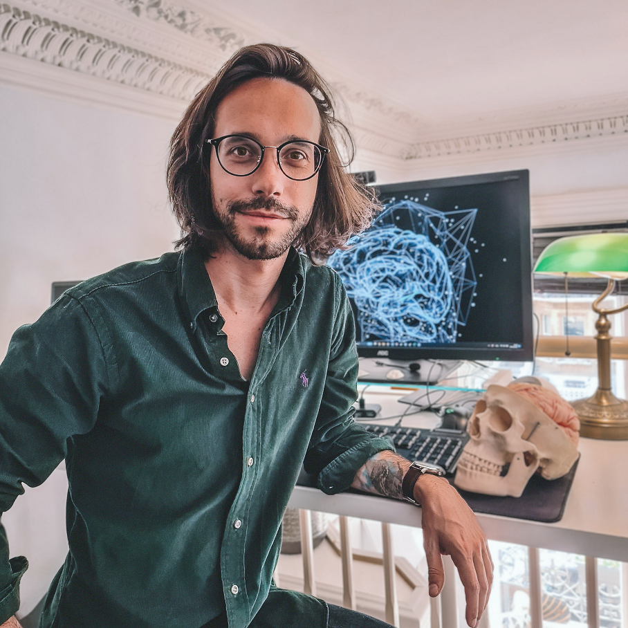 Dominique Makowski, Univ, Sussex (Open science champion)
Dr Dominique Makowski was initially trained as a clinical neuropsychologist and cognitive-behavioural therapist in France, and earned his Ph.D. in psychology from the University of Paris, followed by a postdoctoral training in Singapore. He is currently a lecturer at the University of Sussex, where he leads the Reality Bending Lab, researching the neuropsychology of the experience of reality (e.g., fake news, visual illusions, altered states of consciousness) and the role of the body and emotions in cognition (e.g., emotion regulation and interoception). He is also the author of several popular open-source software, including NeuroKit (for physiological data analysis in Python), easystats (collection of R packages) and Pyllusion (to manipulate visual illusions).
 Hannah Hope, Wellcome (Funder dual perspective)
Hannah Hope is the Open Research Lead within the Research Environment team at Wellcome. Her work cuts across Wellcome’s strategy, supporting Wellcome’s ambition that the research it funds and the processes by which it does this are open, engaged, ethical and efficient. She is responsible for a portfolio of research funding policies and infrastructure grants that aim to create a more open research ecosystem. In the last year, Hannah has been involved in a project exploring tooling to enable research to be more sustainable.
Since her PhD in cell biology, Hannah has been interested in how science is communicated and for what purposes. She worked in scholarly publishing and public engagement prior to joining Wellcome.
Training session: Data Visualisation: Making the most of your data
 Matt Castle, University of Cambridge
This interactive session will explore the principles of effective data visualisation and allow participants to better design and evaluate data visualisations in a range of contexts. We’ll provide guidance for how to critique figures more systematically as well as including an opportunity for participants to put this into practice during the session. No preparation is necessary, and all interaction is voluntary (well it has to be for a session at the end of the day), and unfortunately due to the online nature of the event I’m afraid you’ll have to provide your own coffee, tea and chocolate biscuits.
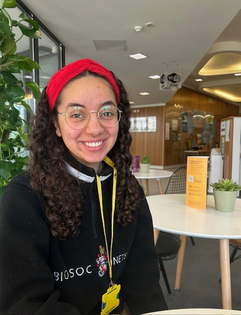 Georgia Boothe, Undergraduate, University of Southampton
Hello! I’m Georgia, an undergraduate integrated masters student in neuroscience, currently studying on my fourth and final year of my degree at the University of Southampton. Within this avenue, I have developed a broad array of skills and explored many facets of the subject, across projects which I have undertaken, including molecular neurobiology, systems neuroscience, and structural neurobiology.
I have also been the student representative for the BNA for past 3 years and continue the pursuit to support neuroscience undergraduates and prospective students from Southampton and across the UK. Notably, this includes various student panels and forums where from a student perspective, our voice can be heard.
I am really looking forward to my future career in science and I hope to strive towards a research-led career in preventative neurodegenerative approaches.
Abstract
Parkinson’s disease (PD) is the 2nd most common neurodegenerative disease following Alzheimer’s disease. In the UK alone there are currently 153,000 living with the condition. There are many factors that contribute to the development of PD such as age, genetics, neuroinflammation, TBI, and exposure to pesticides. Neuroinflammation seems to play a critical role in the early stages of PD.
It was recently found that following TBI, the pro-inflammatory protein S100A9 is a prominent driver in amyloid plaque formation in Alzheimer’s disease. S100A9 is a Ca2+ binding protein that’s main role is to modulate the immune response. The role S100A9 seems to play in Parkinson’s has yet to be resolved.
Through ThT assays and NMR, my research project aims to demonstrate the effects S100A9 has on alpha-synuclein fibrillization. Additionally, it aims to demonstrate the co-aggregation of S100A9 and alpha-synuclein and the different conformational states they take on within this.
If S100A9 was shown to accelerate the rate of α-synuclein forming fibrils, then it would promote further research into how identification of this protein could be a useful diagnostic tool and treatment target for PD.
My talk will cover my project findings and future prospects for this protein.
PANEL SESSION: Molecular, functional and morphological diversity of astrocytes in health and disease
 Valentina Mosienko, Mid-career researcher/clinician, University of Bristol
Dr Valentina Mosienko is an MRC Fellow and Lecturer in Neuroscience at the University of Bristol, at the School of Physiology, Pharmacology and Neuroscience. In the Glia and Psychiatry research group led by Dr Mosienko, they investigate causes, mechanisms and therapeutic targets in neuropsychiatric disorders. The current focus of her research is on non-neuronal mechanisms underlying depression through immune cells of the brain, astrocytes and microglia. Dr Mosienko is passionate about building community of researchers interested in all aspects of glia biology in health and disease – together with other researchers Dr Mosienko recently founded a UK Glia Network.
 Dr Philip Hasel, Mid-Career Researcher (Group Leader and Wellcome Trust Career Development Fellow), UK Dementia Research Institute, University of Edinburgh
Dr Philip Hasel has a PhD from The University of Edinburgh where he studied neuron-to-astrocyte signaling before moving to New York for his Postdoc at the New York University. There, Dr Hasel used single cell RNA-sequencing and spatial transcriptomics to uncover astrocyte subtypes and their unique responses to neuroinflammation. He is now is a Wellcome Trust Career Development Fellow and Group Leader at Edinburgh University studying the role of astrocytes at the borders of the brain and spinal cord.
Talk title: Revisiting the glia limitans in the age of single cell and spatial transcriptomics
 Charlie Friend, Early Career Researcher (Master’s student), University of Bristol
Charlie Friend is a Neuroscience BSc student at the University of Exeter who is currently undertaking a placement in Glia and Psychiatry Lab led by Dr Valentina Mosienko at the University of Bristol. Charlie is actively involved in the project aimed to investigate regulation of astrocyte morphology and number by serotonin. With a foundation in Neuroscience from Exeter, Charlie brings a solid understanding of the field to his current role.
Talk title: Regulation of astrocyte morphology by serotonin
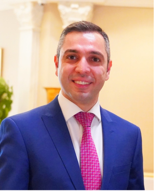 Dr Mootaz Salman, Mid-Career Researcher (Group Leader and MRC Career Development Fellow), University of Oxford
Dr Mootaz Salman is a Group Leader in Cellular Neuroscience and an MRC Career Development Fellow at the University of Oxford. His group aims to investigate mechanisms of blood-brain barrier (dys)function in neurodegenerative diseases and brain injuries using patient-derived stem cells. They design and build innovative dynamic 3D multicellular in vitro models to accurately recapitulate the brain and blood-brain barrier function under mechanobiological stimuli, neuroinflammation and other neurodegenerative-relevant conditions. Dr Salman was awarded the International Society of Neurochemistry (ISN) and APSN Young Neuroscientist Lectureship Award in 2022, The Society for Experimental Biology (SEB) 2024 President’s Medal for the Cell Biology Section and is named the Alzheimer’s Research UK David Hague Early Career Investigator of the Year in 2024.
Talk title: New insights into the regulation of the CNS water homeostasis through targeting aquaporin-4 in astrocytes: from cell biology to clinical trials
Abstract
Brain disorders including neurodegenerative and mental health conditions have tremendous impact on society. Despite dedicated research into the intricate mechanisms of neurological disorders, the efficacy of existing pharmacological treatments is limited, addressing only a select few. Historically, research has predominantly focused on the role of neurons in brain pathologies. Yet, the brain is composed of a vast population of non-neuronal glial cells, with astrocytes being the most abundant among them. Astrocytes play a pivotal role in various brain functions, from regulating development to shaping brain circuits and influencing behaviour. It is now apparent that, similar to neurons, astrocytes can be divided into highly specialized subtypes in both the healthy and diseased brain. Due to their specialization and unique molecular make-up, astrocyte subtypes have become attractive putative therapeutic targets. This symposium brings together researchers aimed to highlight molecular, functional and morphological diversity of astrocytes across the brain and how manipulating these cells can enhance brain functions, potentially offering avenues for preventing and treating brain disorders.
 Ryan Bevan, Research Associate, UKDRI at Cardiff University
Dr. Ryan J. Bevan, a Research Associate at the UK Dementia Research Institute at Cardiff University, is recognised for his contributions to neuroinflammation and neurodegeneration research, with a particular focus on the innate immune system. Currently, Dr. Bevan's research is centred on examining how Alzheimer's Disease microglial risk genes affect pathology in in vivo models. His recent work utilises specialised polychromatic labelling and pathological imaging analysis techniques. Dr. Bevan's future endeavours aim to delve deeper into the role of the innate immune system and microglia involvement in disease pathogenesis, striving to make substantial contributions towards understanding and treating Alzheimer's Disease.
Abstract
A rare protein-coding missense variant (rs72824905) in the PLCG2 gene, which encodes the PLCγ2 enzyme, has been identified as reducing the risk of Alzheimer’s disease. Given its high expression in microglia and position downstream of TREM2, understanding how PLCγ2 is involved in disease mechanisms could reveal its potential as a therapeutic target. We examined the hippocampus and cortex of 6-month-old male and female wildtype and AppNL-G-F mice, carrying either the protective R522 or the risk P522 variants. Mice expressing Plcg2R522 displayed increased microglia densities, less extensive tissue ramifications, and a higher prevalence of the lysosomal marker CD68 compared to those with Plcg2P522. In AppNL-G-F mice, the R522 protective variant counterintuitively increased amyloid plaque core load. These plaques also showed reduced peri-plaque microglia immunoreactivity and lysosomal activity (CD68) compared to the P522 variant. While AppNL-G-F mice experience synaptic loss, those with the R522 variant of PLCγ2 exhibited notable preservation of synaptic integrity in CA1 hippocampal neurons and reduced peri-plaque microglial engulfment of synapses than Plcg2P522 mice. Our findings underscore the role of PLCγ2 in mediating APP-induced synaptic loss, advocating for its consideration as an independent therapeutic target and a complement to amyloid-focused therapies.
Mis-spliced transcripts generate de novo proteins in TDP-43-related ALS/FTD
 Sahba Seddighi, Undergraduate/pre-clinical, University of Oxford
Sahba Seddighi is a neuroscience D.Phil. student at the University of Oxford and a research fellow at the U.S. National Institutes of Health, under the dual mentorship of Prof. Cornelia van Duijn (Oxford) and Dr. Michael Ward (NIH). Her research bridges insights from cellular models of disease and patient biospecimens to investigate the molecular signatures of TDP-43 loss and dysfunction in neurodegenerative diseases, such as amyotrophic lateral sclerosis and frontotemporal dementia. Sahba received her B.A. in neuroscience from the University of Tennessee in 2016 and subsequently completed an M.Phil. in Epidemiology as a Gates Cambridge Scholar at the University of Cambridge before enrolling in medical school at the Johns Hopkins School of Medicine. She intends to pursue a career as a physician-scientist working to make a positive impact in the lives of those living with neurodegenerative diseases.
Abstract
Functional loss of TDP-43, an RNA-binding protein genetically and pathologically linked to ALS and FTD, leads to inclusion of cryptic exons in hundreds of transcripts during disease. Cryptic exons can promote degradation of affected transcripts, deleteriously altering cellular function through loss-of-function mechanisms. However, the possibility of de novo protein synthesis from cryptic exon transcripts has not been explored. Here, we show that mRNA transcripts harboring cryptic exons generate de novo proteins both in TDP-43 deficient cellular models and in disease. Using coordinated transcriptomic and proteomic studies of TDP-43 depleted iPSC-derived neurons, we identified numerous peptides that mapped to cryptic exons. Cryptic exons identified in iPSC models were highly predictive of cryptic exons expressed in brains of patients with TDP-43 proteinopathy, including cryptic transcripts that generated de novo proteins. We discovered that inclusion of cryptic peptide sequences in proteins altered their interactions with other proteins, thereby likely altering their function. Finally, we showed that these de novo peptides were present in CSF from patients with ALS. The demonstration of cryptic exon translation suggests new mechanisms for ALS pathophysiology downstream of TDP-43 dysfunction and may provide a strategy for novel biomarker development.
Cell Type Specific Transcriptomic Signatures of Brain Ageing
 James Tomkins, Research Associate, University of Cambridge
James is a research associate at the Centre for Misfolding Diseases (Department of Chemistry, University of Cambridge) predominantly working on single cell transcriptomics studies of post-mortem human brain, specifically relating to ageing and Parkinson’s disease. He studied BSc Biology at Liverpool John Moores University, MSc Molecular Medicine at the University of Sheffield and was awarded a PhD from the University of Reading in 2020 for research focussed on protein interaction network and C. elegans models of DAPK1 and related proteins.
Abstract
Ageing is the primary risk factor for many neurodegenerative disorders. Despite considerable advances in understanding cellular ageing processes, how these molecular alterations map to specific brain cell types across the lifespan is poorly characterised. Since selective vulnerability of brain regions and cell types is a common phenomenon in neurodegenerative disorders, defining cell type specific hallmarks of ageing at the molecular level is a critical step in understanding the interplay between healthy ageing and progression towards neurodegeneration.
Utilising snRNA-seq data derived from the prefrontal cortex of neurologically healthy donors from across the lifespan post mortem, this project aims to characterise age related molecular signatures for 10 major brain cell types. Data were processed through a standardised pipeline of QC, genome mapping and transcript quantification for individual cell nuclei. Multiple canonical cell type markers were then used to define cell type in our datasets. In total, 94 samples were categorised into 5 age groups spanning human lifespan and cell type specific transcriptomic profiles for each sample were determined. Using this categorisation, comparative analyses are ongoing, such as differential expression and coexpression network analyses. Functional inference will enable these age-associated transcriptomic dynamics to be contextualised in terms of cell type specific biological pathways.
Targeting the infiltrative margin of glioblastomas
 Riddhi Sharma, Early Career, UK Health Security Agency
Hello, my name’s Riddhi! I am currently working as a Neurobiologist, employed at the UK Health Security Agency. I am also completing a part-time PhD in collaboration with the University of Nottingham. My research focuses on glioblastomas, the most aggressive brain tumour, and how we can target them. The primary focus of my PhD is looking at how we can prevent recurrence of these fatal tumours, to help improve patient survival. My talk will discuss several aspects of glioblastomas, including their infiltrative edge, their metabolism, as well as their microenvironment.
Abstract
Gliomas account for between 30-35% of all brain tumours and isocitrate dehydrogenase wild-type glioblastomas, the highest grade of glioma, have a median survival of just 14-16 months from diagnosis. These tumours are incurable and inevitably recur, largely due to the intra- and inter-tumour heterogeneity, which manifests as the tumour progresses. Glioblastomas often develop intrinsic resistance to available standard-of-care treatment, such as concomitant chemotherapy (temozolomide) and radiotherapy, contributing to the 90% recurrence rate associated with glioblastomas. Whilst radiation therapy is a mainstay of standard-of-care, there is a research gap to identify potential synergistic approaches between radiation and molecular targeted therapy, to prevent or substantially delay tumour recurrence. My research will produce novel insights into the recurrence of glioblastomas, by studying cells derived from the primary and recurrent tumour from the same patients. This area has been considerably underexplored, and investigations into the aetiology of the primary and recurrent tumours will shed light on the devastating recurrence and incurability of glioblastomas.
How to develop safe and responsible AI in neuroscience research.
 Lewis Hotchkiss, Early Career, Dementias Platform UK
Lewis Hotchkiss is the Neuroimaging & AI Research Officer at Dementias Platform UK (DPUK) where he leads the development of responsible AI in neurodegenerative diseases.
Abstract
Dementias Platform UK enables data sharing of sensitive and protected healthcare data of patients within a secure environment. This has enabled AI development on these large datasets to understand neurodegenerative diseases and psychiatric disorders which could eventually lead to new tools in diagnosis, prognosis, and treatment in clinical settings. However, there are concerns revolving around the use of AI which utilises protected healthcare data. In our recent research, we aimed to host several workshops with members of the public, researchers and data providers to understand the risks of AI and how we can protect against them when training and developing AI models. From this, we are developing guidelines and recommendations to support the development of safe AI which preserves patient privacy. AI should not only be private, but also ethical, unbiased, and robust. At the Dementias Platform UK Data Portal, we have been finding ways of overcoming all of these barriers to developing safe AI to help researchers be responsible in their research.
Embodied Foraging: Explore vs Exploit social decision making with Japanese Macaques
 Hildelith Leyser, Postgraduate, McGill University
Hildelith’s work focuses on the neural mechanisms of social decision making from an embodied perspective as a Trainee at the RIKEN Center for Brain Science in Japan in the Imagination and Executive Functions Lab (PI: Kentaro Miyamoto). Following an unconventional trajectory, she completed her Bachelor’s degree at the University of Oxford, in History in 2021, and after experiences in humanitarian aid work then went on to complete her Master’s Degree in Applied Neuroscience at the University of London, at Royal Holloway, in 2023. Hildelith is excited about combining ultra-high field fMRI, Transcranial Ultrasound Stimulation and machine learning analysis tools like DeepLabCut to understand the interplay between cognitive functions and motor movements to shed light on how sensory experiences influence our perception of the world and interactions with others.
Abstract
Exploration-exploitation dilemma is characterised as foraging and has been studied from many different approaches in the psychological and cognitive sciences. Most previous research on foraging is conducted using computerised touchscreen-based tasks. However, we hypothesized an overlooked link between movement behaviour and decision-making characteristics due to the overlap between movement planning and decision making neural networks. We devised a 3D foraging paradigm with unknown and known rewards for three Japanese Macaque Monkeys in an unrestrained environment and conducted both individual and social decision-making experiments in this context. We combined analysis of movement patterns with DeepLabCut, a markerless motion pose algorithm, along with models of choice behaviour to shed light on the interconnectedness of movement and social cognition. Our findings showed that exploration and exploitation are characterised by distinct body postures. In addition, optimality and suboptimality of decision making was also reflected in distinctive movement features. Meanwhile we demonstrated that social hierarchy influenced both decisions making strategy and movement patterns. Our findings both complement and complicate established literature on explore/exploit decision-making. These insights have implications for developing embodied therapeutic approaches.
Elucidating the significance of hippocampal perineuronal nets in cognition and behaviour
 Jacob Juty, PhD student, University of Nottingham
I am a BBSRC-funded second year PhD student in behavioural neuroscience at the University of Nottingham, interested in understanding the neural mechanisms underlying cognition in health and neuropsychiatric disease. I am currently researching the role of perineuronal nets in cognition and behaviour, under the supervision of Dr Tobias Bast. In 2022, I graduated from the University of Manchester with a first-class honours integrated masters degree in cognitive neuroscience and psychology.
Abstract
Perineuronal nets (PNNs) have been suggested to?regulate?synaptic plasticity and support?GABAergic inhibition, and PNN disruption has been implicated in brain disorders, including schizophrenia. In the hippocampus, PNNs preferentially surround GABAergic inhibitory neurons. The?cognitive and behavioural consequences of hippocampal PNN disruption largely remain?to be elucidated. PNNs can be experimentally disrupted by chondroitinase-ABC (chABC), an enzyme disrupting chondroitin-sulfate proteoglycans (CSPGs), a main component of PNNs and the diffuse extracellular matrix (ECM). Our aim is to determine how hippocampal PNN disruption, via chABC infusion, affects cognition and behaviour in rats.
As a first step, we characterised hippocampal PNN expression in male Lister hooded rats and the impact of chABC infusion into ventral hippocampus. Wisteria floribunda agglutinin staining revealed PNNs mainly?in CA2/CA3 of dorsal hippocampus and CA1 of ventral hippocampus. ChABC-treated rats showed PNN ablation, alongside attenuation of CSPGs in the diffuse ECM, and C4S “stubs” (products of CSPG degradation by chABC) in ventral to intermediate hippocampus. 10 days after chABC infusion, PNN?staining had partially recovered,?whereas?CSPG staining in the diffuse ECM?remained attenuated.
Our future work will assess the impact of hippocampal chABC infusion on cognitive and behavioural functions linked to hippocampus and regional GABAergic inhibition.
Reducing the carbon footprint of computing in neuroscience: Insights from human neuroimaging
 Nick Souter, Postdoctoral Research Fellow, University of Sussex
Nick is a postdoctoral research fellow within the School of Psychology, at the University of Sussex. He has a background in Psychology and Cognitive Neuroscience, with previous work focusing on the retrieval of semantic concepts, in stroke patients with aphasia and in neurologically healthy adults. Nick’s current work focuses on measuring and reducing the carbon footprint of computing required in human neuroimaging research, with a specific focus on functional magnetic resonance imaging (fMRI). This involves generating evidence-based recommendations for how neuroimagers can reduce carbon emissions resulting from specific digital pipelines - such as FSL, SPM, and fMRIPrep.
Abstract
Research computing has a carbon footprint, and therefore contributes to the climate crisis. This is relevant to neuroscience research, which frequently relies on computationally expensive data processing with large datasets. Fortunately, it is now possible to estimate the carbon emissions of computing, and therefore to reduce them. I will report the findings of a study focused on measuring and reducing the carbon footprint of fMRI preprocessing tool fMRIPrep, using an existing open-source dataset. This will include actionable recommendations as to how neuroimagers can reduce the footprint of their preprocessing without compromising data quality, as measured by statistical sensitivity and data smoothness. For example, disabling FreeSurfer surface reconstruction can reduce estimated carbon emissions by 48%. Conversely, increasing volumetric output space resolution can increase emissions by an alarming 70% with minimal performance gains. I will also share recent results from our follow-up study, in which we are quantifying and comparing the carbon footprint of a wider range of fMRI analysis packages, including FSL, SPM, and fMRIPrep. Finally, I will provide a broader set of recommendations as to how neuroscience researchers can reduce the carbon footprint of their research computing, targeting research practices from study planning through to data sharing.